October 1st, 2009 by Medgadget in Better Health Network, News
No Comments »


An international team of collaborators from a number of academic institutions and a couple pharmaceutical firms has been working with researchers at the National Institute of Standards and Technology (NIST) to study how special sugar coated iron oxide nanoparticles interact with each other to destroy cancer cells under laboratory conditions. The 100 nanometer wide particles, which are attracted by tumor cells, are particularly prone to magnetically induced heating.
Read more »
*This blog post was originally published at Medgadget*
August 28th, 2009 by Berci in Better Health Network, Humor, True Stories
No Comments »

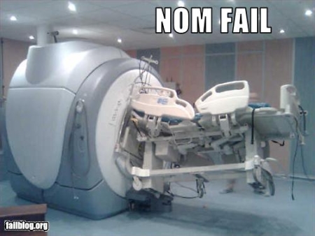
I’ve come across this image on Fail Blog. Magnetic Resonance Imaging + beds with ferromagnetic parts equal…

*This blog post was originally published at ScienceRoll*
July 30th, 2009 by Medgadget in Better Health Network, News
No Comments »

 The scans presented here are of a ten year-old German girl who was discovered to be missing the right hemisphere of her brain. Incredibly, she is perfectly normal, except for a history of seizures and a slight weakness on her left side. Attending school with others of her age, it is reported that she is able to study and play sports, just like other kids around her. Of course, the mystery is how is this all possible? To answer the question, University of Glasgow scientists used an fMRI to see where the left eye’s vision is processed. Turns out that the brain’s visual area responsible for the right eye offered up some space for the left.
Normally, the left and right fields of vision are processed and mapped by opposite sides of the brain, but scans on the German girl showed that retinal nerve fibres that should go to the right hemisphere of the brain diverted to the left.
Further, the researchers found that within the visual cortex of the left hemisphere, which creates an internal map of the right field of vision, ‘islands’ had been formed within it to specifically deal with, and map out, the left visual field in the absence of the right hemisphere.
Dr Lars Muckli of the Centre for Cognitive Neuroimaging in the Department of Psychology, who led the study, said: “This study has revealed the surprising flexibility of the brain when it comes to self-organising mechanisms for forming visual maps.
“The brain has amazing plasticity but we were quite astonished to see just how well the single hemisphere of the brain in this girl has adapted to compensate for the missing half.
“Despite lacking one hemisphere, the girl has normal psychological function and is perfectly capable of living a normal and fulfilling life. She is witty, charming and intelligent.”
The girl’s underdeveloped brain was discovered when, aged three, she underwent an MRI scan after suffering seizures of brief involuntary twitching on her left side.
The scientists believe the right hemisphere of the girl’s brain stopped developing early in the womb and that when the developing optic nerves reached the optic chiasma, the chemical cues that would normally guide the left eye nasal retinal nerve to the right hemisphere were no longer present and so the nerve was drawn to the left.
This implies that there are no molecular repressors to prevent nasal retinal nerve fibres from entering the same hemisphere.
Dr Muckli added: “If we could understand the powerful algorithms the brain uses to rewire itself and extract those algorithms together with the general algorithms that the brain uses to process information, they could be applied to computers and could result in a huge advance in artificial intelligence.”
Press release: Scientists reveal secret of girl with ‘all seeing eye’…
*This blog post was originally published at Medgadget*
November 24th, 2008 by Dr. Val Jones in Audio, News
1 Comment »
I really like new technology, especially when it offers a very obvious advantage for patients. I recently heard about a new CT scanner that is so fast, it dramatically reduces radiation exposure for patients and can take crisp images of moving organs (like the heart). I asked to speak with Siemens’ VP of Sales and Marketing, Dr. André Hartung, to find out about the new Somatom Definition Flash Dual Source CT Scanner (it takes longer to say the machine’s name than to scan your entire body). Of course, I invited my Medgadget friend, Gene Ostrovsky, to join the call. I’ve included a “bonus track” for more advanced readers at the end of this blog post. Enjoy!
Listen to the podcast here:
[audio:http://blog.getbetterhealth.com/wp-content/uploads/2008/11/andrehartunglowq1.mp3]
Dr. Val: Just to set the stage for our listeners – can you explain what a CT scanner is, and how it differs from an MRI?
Hartung: Both CT scanners and MRI machines allow healthcare professionals to look inside the human body for diagnostic purposes. While CT scanners use x-rays to produce images, MRI machines use magnets. CT Scanners are very fast and widely available – almost every hospital has one.
Dr. Val: When would a doctor want to use a CT scanner instead of an MRI machine?
Hartung: CT images are especially good at detecting cancer. Also, because CT scans can be done so quickly, they are also useful diagnostic tools for stroke, heart attack, or when a patient is in critical condition – when every second counts.
Dr. Val: You said that CT scans are based on x-ray technology. How much radiation exposure does the average CT scan cause?
Read more »


 The scans presented here are of a ten year-old German girl who was discovered to be missing the right hemisphere of her brain. Incredibly, she is perfectly normal, except for a history of seizures and a slight weakness on her left side. Attending school with others of her age, it is reported that she is able to study and play sports, just like other kids around her. Of course, the mystery is how is this all possible? To answer the question, University of Glasgow scientists used an fMRI to see where the left eye’s vision is processed. Turns out that the brain’s visual area responsible for the right eye offered up some space for the left.


