December 15th, 2009 by Shadowfax in Better Health Network, Humor, True Stories
No Comments »

 They’re not allowed to actually write “Hey Dummy, look here” on the x-ray report, but this is what the radiologists do when they want to make sure the idiots in the ER won’t miss the key finding on a film (in this case, a bit of glass from an automobile window):
The wonders of digital radiography allow this to appear on my computer screen. In the old days they did it with a grease pencil and a post-it note.

*This blog post was originally published at Movin' Meat*
June 26th, 2009 by scanman in Better Health Network, True Stories
2 Comments »

…
I was called to do an urgent bedside ultrasound scan of the abdomen for a trauma victim.
The patient was a young man of twenty-four who had been involved in a road traffic accident (RTA = MVA in US medical terminology). He had been brought – without any kind of basic life support – after sustaining a major trauma at a village about two hours away. The intensivist in the ICU told me that he was in severe hypovolemic shock on admission with a GCS of 4. Preliminary examination and radiographs had shown a comminuted fracture of the right femur (thigh bone) with a large hematoma and some facial bone fractures. After initial assessment and resuscitation in the casualty, a CT scan was done. He had a fracture in the frontal bone and a few small contusions in the brain, that raised the possibility of Diffuse Axonal Injury, nothing that could explain a GCS of 4 though. The assumption was that it was all due to extensive blood loss and hypovolemia. He was shifted straight to the ICU after the CT scan and I was called to do an ultrasound scan to check for hemoperitoneum (ie, abdominal injury and blood loss).
The scan was normal. As I was doing the scan, the intensivist was busy trying to put in a Subclavian central line. He secured the line just as I finished my scan, which incidentally was normal. As I was stepping away from the bed, the patient had a cardiac arrest, as evidenced by sudden bradycardia on the monitor. I moved out of the way as the intensivist, orthopaedic surgeon and ICU nurses went through a full resuscitation protocol. After a while, even I realized that it seemed like a futile exercise.
I was not particularly busy, so I peeped into to the Cardiac ICU next door as there seemed to be some commotion there. My cardiologist colleague, a normally friendly soul was peering intently at a very fast heart rhythm on a monitor over the bed of a young girl of about six or seven. There were a couple of nurses injecting something slowly into an intravenous cannula in the kid’s forearm. In passing, I noted that the kid was very calm and seemed very interested in what the nurse was doing. I stepped close to my friend and asked what was up. He turned, gave me a quick nervous smile and said he was trying to revert an SVT (supraventricular tachycardia, a very nasty fast heart rhythm). Honestly, I had never seen an SVT in someone so young, so I asked him what was the history. He told me the kid was brought by her mother to his outpatient clinic a short while ago because she complained of palpitations (I forgot the exact description used by the kid, but it was quite descriptive). My friend said he was sure it was an SVT after a quick examination in the clinic, so he rushed the kid upstairs to the Cardiac ICU, connected her to a monitor, confirmed the diagnosis and had ordered Adenosine IV stat for reversal. He maintained his intent survey of the monitor as he recounted the story and the nurse continued her slow IV injection. At one particular point when the line on the monitor became particularly squiggly, he shouted, “STOP!” and the nurse stopped injecting.
…
It was almost magical.
…
The squiggles became a recognizable cardiac rhythm, albeit very fast – about 160 to 170 beats per minute. My friend called out to one of the superfluous nursing attendants and asked them to get the kid’s mother inside. A very anxious young lady who had obviously been weeping was led in. My friend showed her the monitor and explained to her that the nasty rhythm had been made to behave itself or something to that effect and told her that the kid was out of any imminent danger.
Happy with the positive outcome, I strolled out to be confronted by a wailing family, including two young girls, maybe a year or two older than the calm kid inside, who had just been told that their older brother who fell off his motorbike was dead.
It was past my work hours. I went out and had a drink and reflected.
Such is life.
…
*This blog post was originally published at scan man's notes*
May 17th, 2009 by Dr. Val Jones in True Stories
3 Comments »
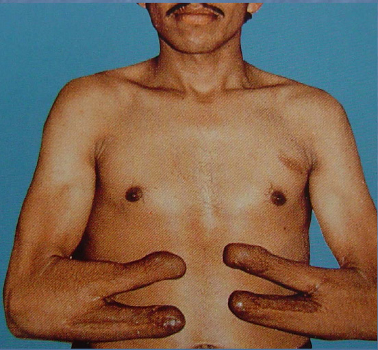
Tragically, land mines injure between 15,000 to 20,000 people each year. Some civilians see a metal object sticking out of the ground and attempt to pick it up and inspect it – the result is often loss of both hands and eyes.
The goal of rehabilitation after trauma is to restore as much independence as possible to patients. With loss of vision and no hands, self care, feeding, and donning/doffing arm prostheses can be very challenging. There is a procedure, known as the Krukenberg operation (named after Hermann Von Krukenberg, who first described it in 1917), that allows the forearm bones to be separated, using the muscle rotators that exist between them to create a pincer grasp. This procedure is not uncommonly used in India and Pakistan and does indeed return some degree of functional use to the arms.
At a recent Physical Medicine and Rehabilitation conference, this photograph was used to illustrate arm function after the Krukenberg operation.

Photo Credit: Dr. Heikki Uustal
It certainly presents a conundrum – should function trump aesthetics in all cases?
I’m not sure that I’d want this procedure, even if I lost my vision and both hands.
Would you?
 They’re not allowed to actually write “Hey Dummy, look here” on the x-ray report, but this is what the radiologists do when they want to make sure the idiots in the ER won’t miss the key finding on a film (in this case, a bit of glass from an automobile window):


