April 1st, 2011 by admin in Book Reviews
No Comments »


Hall WA, Nimsky C, Truwit CL. Intraoperative MRI-Guided Neurosurgery. Thieme 2010, 272 pages, $159.95.
This book is a multiauthored text edited by three senior authors who have a tremendous experience in the use of intraoperative MRI technology. The book is divided into five sections that describe the various iterations of iMRIs that are available, its application for minor procedures, the resection of neoplastic lesions, and its role in the management of nonneoplastic disorders. The last section focuses on the future improvements in design that are likely to improve surgical access and utility of this burgeoning technology.
The first section describes the characteristics of iMRI machines that are available in the low, medium and high field strength. The reader gets a very good idea about the relative benefits and limitations of each of these machines. Hospitals that may be in the process of deciding which technology to go in for may use this information as a good guide. This section also highlights the optimal pulse sequences that may help differentiate tumor-brain interface, perform intraoperative fMRI and DTI tracking and detect complications related to brain ischemia and hematoma formation. The chapters in this section are well illustrated and show both the technology and the images obtained with various units. The chapter on optimal pulse sequences is very well written and discusses the specific pulse sequences that can help obtain the maximum intraoperative information with the least amount of time. These sequences can be tailored to provide not only anatomical details but also to help obtain both DTI and functional activation data for intraoperative neuronavigation, thereby accounting for brain shifts and movement of eloquent tracts during surgery. The authors describe the challenges of this methodology. Specific anesthetic challenges that restrict the use of standard monitoring equipment have been outlined. These include patient access, length of operative procedure, influence of magnetic field and RF currents on the functioning of the equipments and the images obtained, and risk of migration of ferromagnetic instruments, among others. This has led to the development of MR compatible anesthesia and monitoring equipment. Safety issues and steps needed to ensure reliability of equipment have been described. Read more »
*This blog post was originally published at AJNR Blog*
March 21st, 2011 by admin in Health Tips, True Stories
5 Comments »

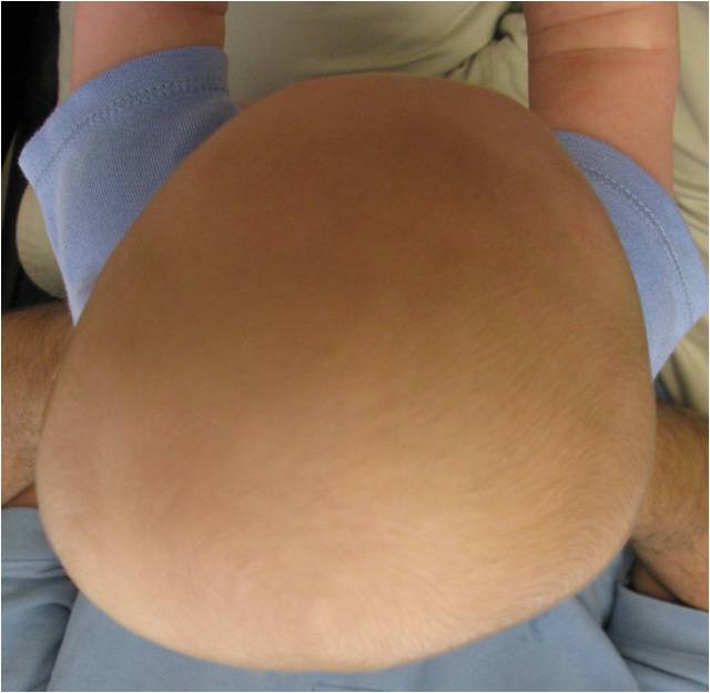
Figure 1
This post was contributed by guest blogger, Edward Ahn, M.D.
The head coach of a Division 1 champion women’s sports team brought her baby daughter in to me for evaluation of her flat head at the recommendation of her pediatrician.
While I was examining her baby, I started to say, “Well, I’ll tell you what she has —
She quickly interrupted, “Is it bad?”
I looked up to see fear written on this tough coach’s face. I was struck by how this benign condition can cause apprehension in so many parents.
Often, pediatric neurosurgeons like me or plastic surgeons are asked to assess babies with a flat head, also known as positional plagiocephaly. Usually, parents have developed a fair amount of anxiety, often with the underlying fear that their baby will need surgery or the brain will grow abnormally. These fears are not warranted. Read more »
March 20th, 2011 by American Journal of Neuroradiology in Research
No Comments »

We report a pathologically proved craniopharyngioma in the prepontine cistern. A 50-year-old woman presented with swallowing difficulty for 1 month. She underwent brain MR and CT imaging.
T1-weighted, T2-weighted, and contrast-enhanced T1-weighted images showed a large peripheral enhancing cystic mass in the prepontine cistern. Inside the lesion, high signal intensity (SI) on T1 and low SI on T2-weighted imaging were noted (Fig 1). The CT scan showed features similar to those on the MR images, except for the addition of a peripheral small calcification in the cystic lesion. We could not find any connection between the mass in the prepontine cistern and the sellar or parasellar area. The mass was partially surgically removed, and histopathologic examination revealed craniopharyngioma in the prepontine cistern.

View larger version (102K):
[in this window]
[in a new window]- Fig 1. A 50-year-old woman with a craniopharyngioma in the prepontine cistern. A, Sagittal T1-weighted image shows a cystic mass in the prepontine cistern. B, Contrast-enhanced T1-weighted sagittal image shows a peripheral enhancing cystic mass in the prepontine cistern. Read more »
*This blog post was originally published at AJNR Blog*
June 24th, 2010 by Medgadget in Better Health Network, News, Research
No Comments »

 Scientists at Arizona State University have developed a new method of non-surgical brain stimulation using pulsed ultrasound that enhances cognitive function in mice, and may one day be used to non-invasively treat patients with mental retardation, Alzheimer’s disease and other central nervous system (CNS) dysfunctions.
In intact motor cortex in mice, ultrasound was found to stimulate action potentials and elicit motor responses comparable to those only previously achieved with implanted electrodes and related techniques. It also activates meaningful brain wave patterns and the production of brain-derived neurotrophic factor (BDNF) in the hippocampus — one of the most potent regulators of brain plasticity. Read more »
*This blog post was originally published at Medgadget*
June 15th, 2010 by Medgadget in Better Health Network, News, Research
No Comments »

 In the continuing effort to make surgery less invasive, physicians at Johns Hopkins Hospital are operating on the brain through a tiny incision in one of the eyelids instead of lifting a large piece of the skull.
Named transpalpebral orbitofrontal craniotomy, the procedure allows for access to the middle and front regions of the brain. The cranial cavity is reached through a hole created by removing a small, half-inch to one-inch-square section of skull bone right above the eyebrow. Endoscopic surgery can then be performed with help of previously obtained CT and MRI data. Read more »
*This blog post was originally published at Medgadget*


 Scientists at Arizona State University have developed a new method of non-surgical brain stimulation using pulsed ultrasound that enhances cognitive function in mice, and may one day be used to non-invasively treat patients with mental retardation, Alzheimer’s disease and other central nervous system (CNS) dysfunctions.
 In the continuing effort to make surgery less invasive, physicians at Johns Hopkins Hospital are operating on the brain through a tiny incision in one of the eyelids instead of lifting a large piece of the skull.


